Patient education guide
Taking the next step in your glaucoma intervention
Understand how glaucoma affects vision and explore the surgical options your eye doctor may recommend to help lower pressure.
Begin the journey
Understanding glaucoma
What is glaucoma?
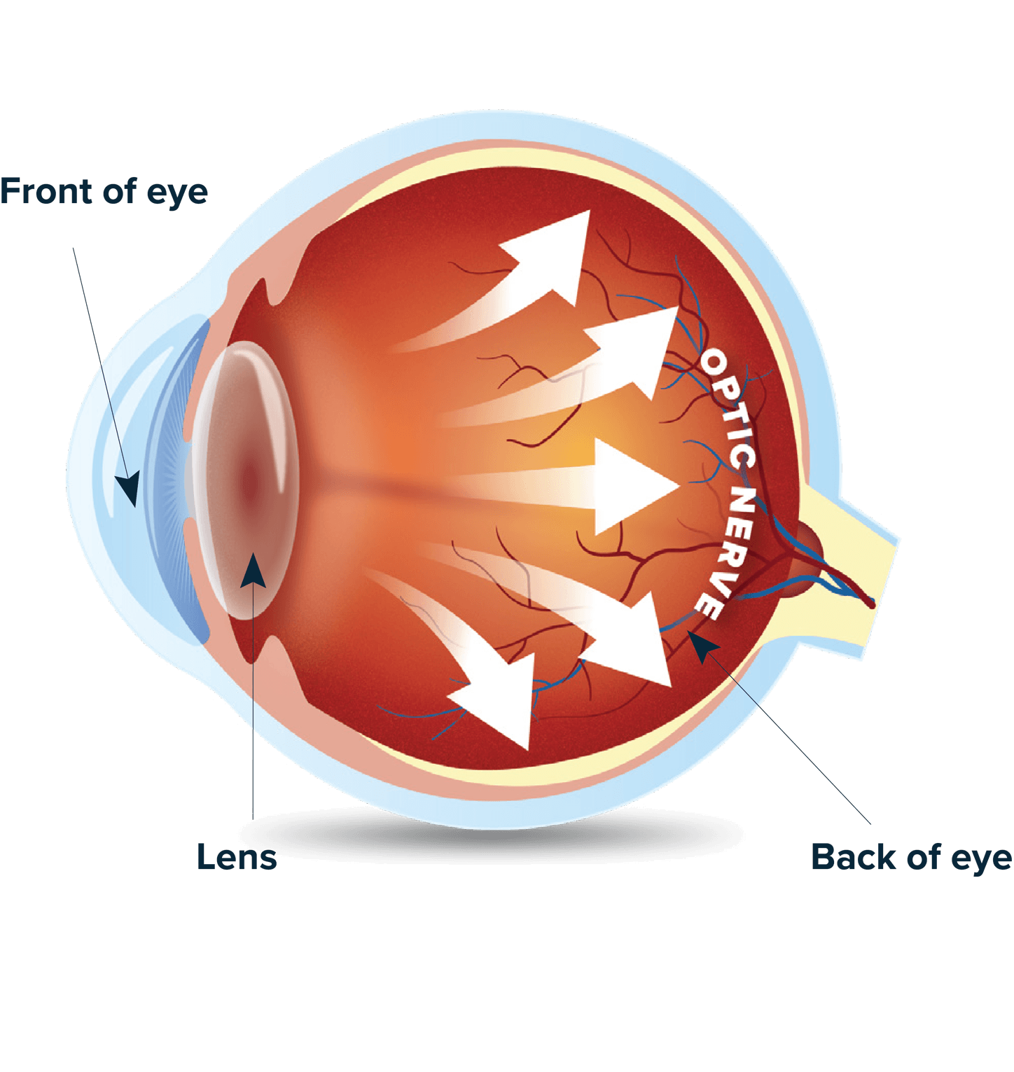
Glaucoma occurs when the optic nerve is damaged, often due to a buildup of fluid pressure in the eye that does not drain properly. That damage can result in permanent vision loss.
Fluid normally leaves the eye through a natural drainage system. If that system becomes blocked or less efficient, pressure can build and push on the optic nerve, which carries visual signals to the brain.
How sight can change
Vision loss with glaucoma
Vision loss tends to begin at the outer edges and may be difficult to notice at first. The images below are illustrative, not a real depiction of glaucoma.



Regular eye exams matter because glaucoma can get worse before you notice symptoms.
Understanding eye drainage
Why are procedures necessary?
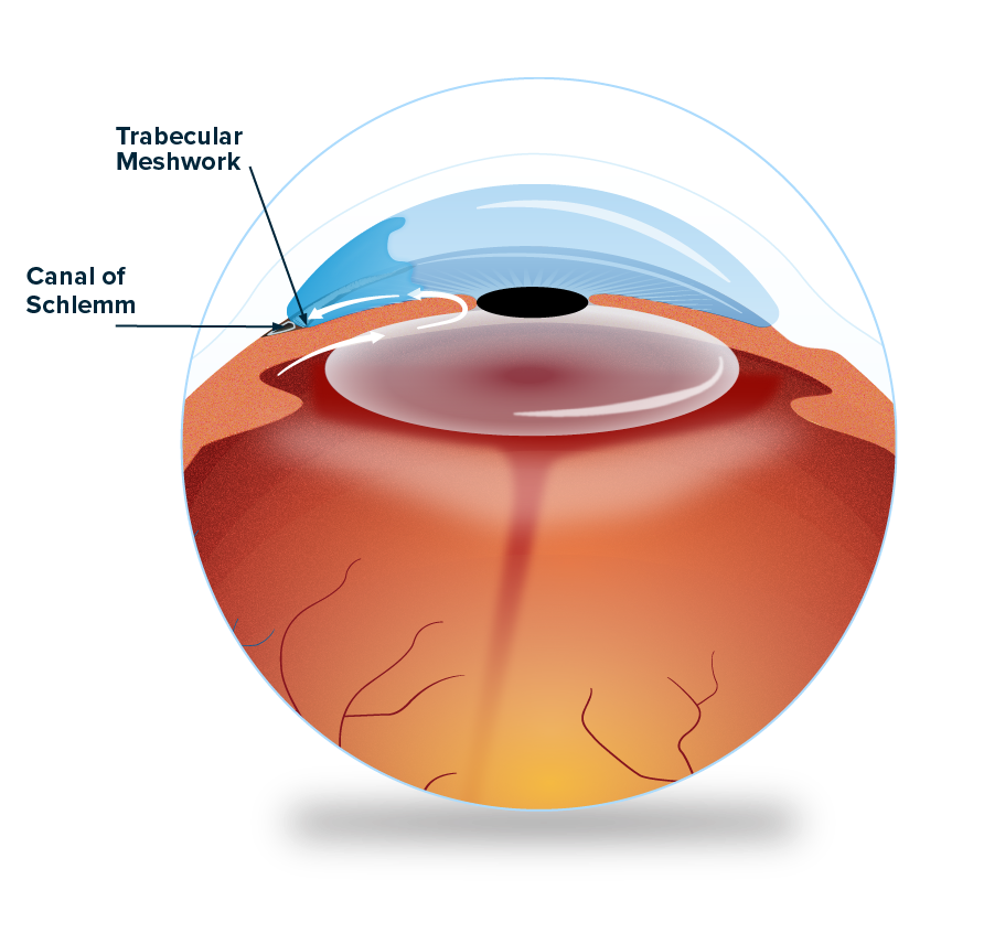
Surrounding your eye is a drainage system that moves fluid away from your eye, like gutters preventing water from pooling up around a house.
Two important structures in that system are called the Schlemm’s canal and Trabecular Meshwork.
- Schlemm’s canal: provides a channel for fluid to leave your eye, but it can become collapsed or blocked. When that happens, fluid will struggle to leave your eye until Schlemm’s canal is fixed or cleared of obstruction.
- Trabecular Meshwork: normally works as a filter or net, providing resistance to fluid leaving your eye and ensuring the correct amount of fluid is allowed to leave. Over time, this net can become stiff or blocked, making it difficult for any fluid to drain properly. When this happens, pressure can remain high even when other treatments have been used.
Flushes the eye’s natural drainage system
Canaloplasty
What is the procedure?
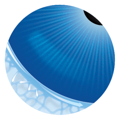
Canaloplasty is a gentle procedure designed to clear obstructions and flush the eye’s natural drainage system. Your surgeon enters Schlemm’s canal with a tiny tube or cannula and injects a soft, gel-like material called viscoelastic.
The material gently expands the canal from within, widens areas that may have narrowed or collapsed, and clears the pathway so fluid can flow more freely. The eye’s natural anatomy is left intact and nothing permanent is left in place. It may be performed by itself or with cataract surgery.
Goals of the procedure
- Improve the eye’s natural drainage
- Lower eye pressure compared with drops alone
- Potentially reduce the number of daily eye drops
- Resume most daily activities in a few days
What to expect after
Slight discomfort in the treated eye may occur. Your doctor will continue to monitor your eye pressure and adjust your treatment as needed.
Removes a small strip of blocked tissue
Goniotomy
What is the procedure?
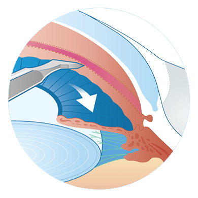
When the trabecular meshwork has become severely blocked or diseased, a goniotomy removes a small strip of that tissue to create a clearer path into the eye’s natural drainage channels.
Some techniques treat one area, while others remove a larger strip to allow more drainage. Goniotomy works with the eye’s natural drainage system, leaves no permanent implant behind, and can be performed by itself or with cataract surgery.
Goals of the procedure
- Improve the eye’s natural drainage
- Lower eye pressure compared with drops alone
- Potentially reduce the number of daily eye drops
- Resume most daily activities in a few days
What to expect after
Slight discomfort in the treated eye may occur. Your doctor will continue to monitor your eye pressure and adjust your treatment as needed.
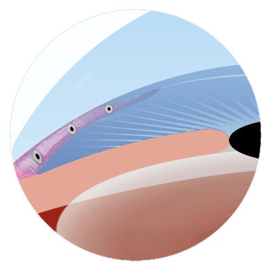
Bypass local blockages
Implantable Stents
What is the procedure?
Stents are small devices placed inside the eye to help direct fluid out more easily. The implant acts like a tiny channel, guiding fluid away from areas of resistance toward a place where it can be absorbed by the body.
Depending on the type, a stent may help fluid move into the eye’s natural drainage channels or direct it to a small area just outside the eye. Stents are placed through a small incision, often with cataract surgery, though sometimes on their own.
Goals of the procedure
- Help fluid drain from the eye
- Lower eye pressure compared with drops alone
- Potentially reduce the number of daily eye drops
- Resume most daily activities in a few days
What to expect after
Slight discomfort in the treated eye may occur. Your doctor will monitor your eye pressure and adjust your treatment as needed.
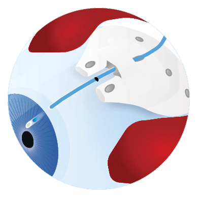
Create an alternative path for fluid to flow
Tube Shunts
What is the procedure?
Tube shunts, also called glaucoma drainage devices, are artificial filtering devices often used when glaucoma is more advanced and less invasive procedures may not provide enough pressure control.
A small tube drains fluid from inside the eye to an attached plate. The plate supports formation of a natural reservoir on the eye’s outer surface, where fluid is gradually absorbed. The tube is covered with tissue and the plate sits beneath the eye’s outer layer, so it is not visible.
Goals of the procedure
- Provide a long-term, substantial reduction in eye pressure
- Create a new pathway when natural drainage is not functioning well
What to expect after
Recovery may take longer than with other procedures. Temporary redness, blurry vision, discomfort, and bleeding immediately after surgery may occur. Extra visits and management may be needed while the natural reservoir forms, and your care team will monitor you closely.